AI for Pulmonary Embolism Detection
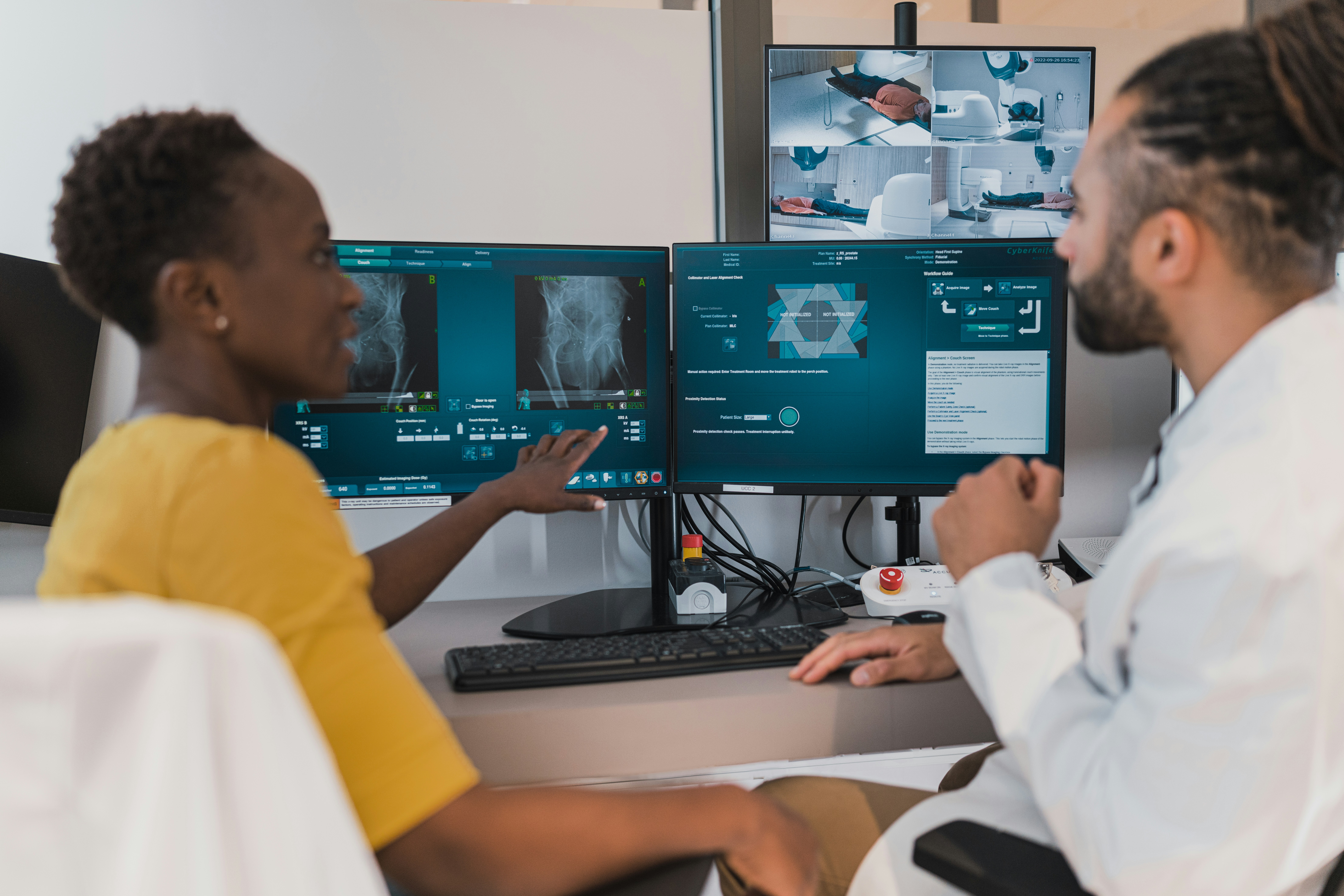
Pulmonary Embolism (PE) diagnosis using machine learning (ML) techniques on CT pulmonary angiography (CTPA) is transforming the field of medical imaging. PE is a life-threatening condition where blood clots block the pulmonary arteries, requiring timely and accurate detection to improve patient outcomes. Traditional training and development of ML models for PE diagnosis involve extensive annotation, which is resource-intensive. To make these applications practical, the team has developed optimized training strategies, model architectures, and deployment pipelines for efficient PE detection.
Read more...Keywords:
Artificial Intelligence (AI), Machine Learning, Biomedical Imaging, Image & Video Processing, Software, Advanced Health Technologies, Point-of-Care, DiagnosticsNovel ML for pulmonary function testing on oscillometry devices
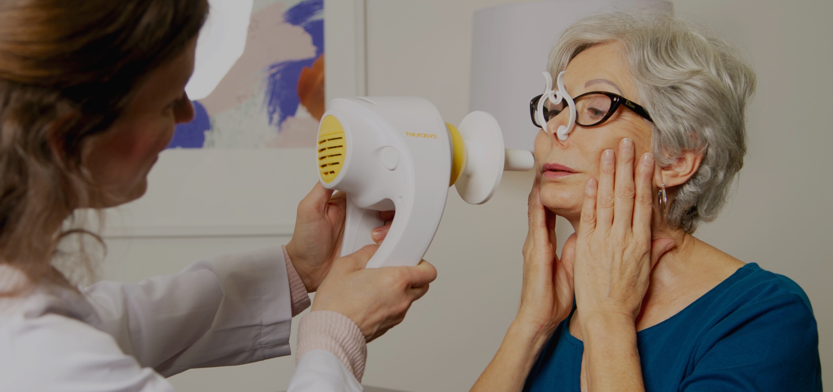
Machine learning architecture offers a novel, high-fidelity means of assessing lung physiology during the performance of (monofrequency) oscillometry tests.
Read more...Keywords:
Machine Learning, Artificial Intelligence (AI), Advanced Health Technologies, Diagnostics, Image & Video Processing, Point-of-Care, Biomedical Imaging, SoftwareRapid, remote vital sign monitoring and screening
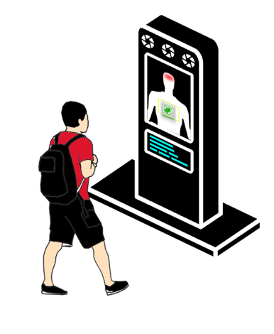
Based on thermal and optical sensing analysis of digital imaging, this technology allows for rapid measurement of photoplethysmogram (PPG), temperature, respiratory rate, and heart rate beyond conventional distance limitations (e.g. at distances more than 1m), with the ability to add other components (e.g. tissue oxygenation and blood pressure) in the future.
Read more...Keywords:
Advanced Health Technologies, Covid-19, Remote Monitoring, Patient Care, Biomedical Imaging, Software, 3D Imaging, Image & Video ProcessingRapid, HiFi MRI Data Processing
A DCE-MRI acquisition and reconstruction package that enables more accurate quantification of physiological parameters in larger volumes than any state-of-the-art method. By first performing a high-temporal (rapid), high spatial (hifi) acquisition of anatomical details in the image set, each time frame is reconstructed with a high degree of precision and accuracy.
Read more...


