BACKGROUND
-
Dynamic contrast-enhanced magnetic resonance imaging (DCE-MRI) is a quantitative method for in vivo tissue examination, where sequential images are taken following administration of a contrast agent. DCE-MRI is useful for:
- Measuring vessel wall leakiness
- Diagnosing and assessing cancer, cardiac health, rheumatoid arthritis, and other diseases
- Regenerative medicine
- Monitoring treatment
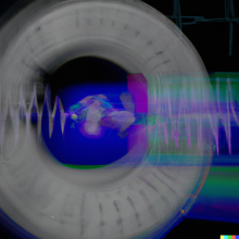
- In order to acquire and reconstruct DCE-MRI images for analysis, state-of-the-art methods (e.g. golden angle radial sparse parallel (GRASP) and its successor, GRASP-PRO) take multiple 1D frames of the same space over time.
- Problem: At higher acceleration rates, these methods fail to capture fine anatomic detail.
TECHNOLOGY OVERVIEW
- Our product is a DCE-MRI acquisition and reconstruction package that enables more accurate quantification of physiological parameters in larger volumes than any state-of-the-art method.
- By first performing a high-temporal (rapid), high spatial (hifi) acquisition of anatomical details in the image set, each time frame is reconstructed with a high degree of precision and accuracy.
- IP has been filed around creating a small number of low temporal resolution image frames with high spatial resolution, which is then augmented to a full reconstruction with high temporal and spatial resolution.
BENEFITS
- Existing efforts attempt to accelerate acquisitions by undersampling, or acquiring less than the full dataset, while at the same time reconstructing images at their full spatial resolution. Other approaches rely on a vast amount of training data for machine learning that may need to be retrained and have the potential to create diagnostically misleading artifacts (“noise”).
- Unlike other approaches, our method can attain much higher acceleration factors without compromising spatial resolution and preserve details without sacrificing acquisition speed.
- Our method is:
- Patient-specific: Works out-of-the-box and does not need to be trained/ retrained on external datasets
- More accurate: Preserves spatial details without sacrificing acquisition speed
- Faster: Attains much higher acceleration without compromising spatial resolution
- Which enables:
- Accurate quantitative and qualitative analysis
- Imaging of larger volumes
- Whole heart DCE-MRI
- Real-time capture of blood flow kinetics
- Reduced repeat examinations
- Scan time reduction
- Faster triage
- Better predictions, diagnosis, detection, and clinical decision making
APPLICATIONS
- Rapid temporal dynamics is necessary for analysis of pharmacokinetics and accurate estimation of physiological parameters such as perfusion. High spatial resolution is necessary for precise delineation of small structural details, which is paramount in medical diagnosis. Because these requirements are difficult to satisfy simultaneously with current imaging techniques, the preference is to retain an acceptable level of spatial resolution at the expense of potentially inaccurate pharmacokinetic estimation.
- The medical imaging and informatics market is set to grow to more than $45 billion globally by 2025, spurred by providers’ investments in solutions that: increase efficiency and productivity, reduce scan times and repeat examinations, and improve triage and diagnoses (Frost & Sullivan, 2021).
- Can help open new applications in perfusion MRI, vasoreactivity MRI (e.g. stress testing), neuro-functional MRI, and CINE MRI.
STATUS
- PCT filed filed September 2023
- TRL 6: Near-final version of the package has been validated in simulated operational environments including on in-vivo data.


Laurent Moreno
Innovations & Entrepreneurship Manager
Innovations & Partnerships Office (IPO)
(416) 946-0594