BACKGROUND
Osteoclasts are large multinucleated cells responsible for bone resorption and calcium regulation in the body. They play a crucial role in the pathogenesis of chronic osteolytic diseases such as periodontitis, osteoporosis, and rheumatoid arthritis. Osteoclastogenesis, the process of osteoclast formation, is mediated by macrophage colony-stimulating factor (M-CSF) and receptor activator of nuclear factor-kB ligand (RANKL). Despite extensive research, the mechanisms that mediate fusion during osteoclastogenesis remain undefined. Actin filaments are especially important for the formation of podosomes, primary adhesive structures, during the fusion process. However, the molecular events that regulate fusion occur only transiently during the brief fusion process, necessitating better tools to visualize and study this fusion during osteoclastogenesis.
TECHNOLOGY
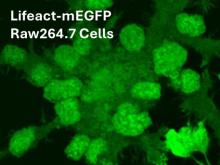
Researchers at the University of Toronto have established a stable Lifeact–mEGFP-transfected RAW264.7 cell line to examine the role of actin filaments in preOC fusion. Lifeact is a peptide that binds to filamentous actin (F-actin) and, when fused to green fluorescent protein (GFP), allows for real-time visualization of actin dynamics in cell expressing this construct (Figure 1). Two distinct cell lines have been produced: H10, which forms large multinucleated osteoclasts, and C8, which does not fuse. The difference in these cells lies in the expression of CD109 which is significantly upregulated in the H10 cell line during osteoclastogenesis, suggesting its critical role in this process.
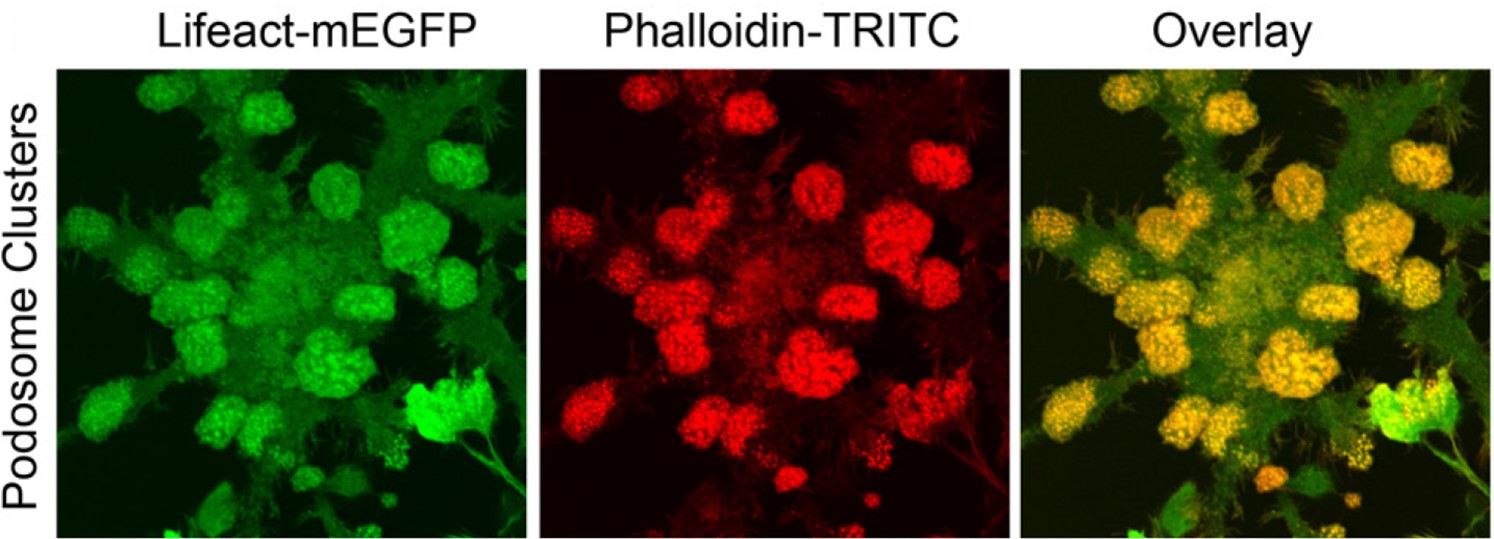
Figure 1. Lifeact–mEGFP enables monitoring of actin filament dynamics in OCs. F-actin identified by Lifeact-mEGFP correlates with that identified by tetramethylrhodamine (TRITC)-conjugated phalloidin.
COMPETITIVE ADVANTAGE
- Enhanced Visualization: LifeAct-GFP cells provide superior visualization of actin dynamics compared to traditional staining methods.
- Real-Time Monitoring: Allows for real-time observation of osteoclast formation and fusion processes.
- Quantitative Analysis: Enables precise quantification of osteoclast formation and fusion efficiency.
APPLICATIONS
- Bone Disease Research: Studying the mechanisms of osteoclastogenesis in diseases such as osteoporosis, periodontitis, and rheumatoid arthritis.
- Drug Development: Screening potential therapeutics that target osteoclast formation and activity.
INTELLECTUAL PROPERTY STATUS
- Proprietary cell lines (H10 and C8)
PROJECT STATUS
Stably transfected LifeAct-GFP RAW264.7 cells have been successfully engineered and characterized. Fursion during osteoclastogenis has been studied using H10 and C8 cells.
KEYWORDS
Bone Resorption, Actin Dynamics, Cell Fusion, Fusopods, Cell Imaging, Bone Disease Research, Cell Lines, Biomedical Research Tools