BACKGROUND
Fibrosis, or scarring of tissue, is not limited to the skin after an injury. In many chronic conditions, such as hypertension, autoimmune lung disease, and alcohol abuse (affecting the heart, lungs, and liver, respectively), tissue becomes progressively fibrotic over time, a condition that often goes undetected but ultimately results in organ failure and even death. Unfortunately, there is no cure for late-stage fibrosis, and tissue biopsy remains the only reliable diagnostic option, despite the deficiencies of limited sampling and mis-sampling. At the University of Toronto, our inventors have developed a non-invasive magnetic resonance imaging (MRI) compound that can be administered to the patient to “light up” fibrotic tissue, including newly developing scars, in 3D throughout the body, to find fibrosis at early stage when the opportunity to interrupt disease progression is the greatest.
TECHNOLOGY
Our inventors have designed a new MRI fibrosis-targeting contrast agent that has an affinity for the substrate of which fibrotic tissue is composed. It is capable of binding to both newly developing scar and older scars. In contrast to other non-invasive imaging methods that claim to image fibrosis, our targeted agent does not highlight dead tissue space – it will highlight only scar tissue, providing a degree of specificity hereto unseen. Imaging on an MRI scanner requires simply a T1-weighted scan, which is widely available on any MRI scanner.

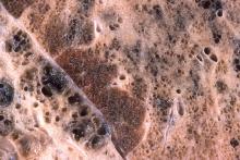
Figure 1. MRI of mouse left ventricle at 4-weeks after gold-standard isoproterenol administration for inducing myocardial fibrosis. Diffuse enhancement of papillary muscle (arrow) and endocardium (chevron) is best seen 10 hours post-fibrosis-targeting agent. Graph of T1 relaxation times shows no change in control hearts but significant reduction in fibrotic hearts
COMPETITIVE ADVANTAGE
- Sensitive and specific detection capabilities
- Unlike other MRI contrast agents, our agent does not contain toxic metals
- Synthesis of new fibrosis-targeting contrast agent is simple and scalable
- Easily incorporated into a clinical contrast-enhanced MRI workflow
- No special imaging protocol required
APPLICATIONS
- Clinical trials for the following indications:
- Cardiac fibrosis from hypertension, diabetes, obesity, and infection
- Liver fibrosis from alcohol abuse, viral infection, and fatty liver disease
- Lung fibrosis from viral/bacterial infection, environmental allergens (e.g. asbestos, dust, etc.), radiation therapy, chemotherapy, and many lung diseases
- Kidney fibrosis from diabetes, hypertension, autoimmune disease, and infection
- Intestinal fibrosis from inflammatory bowel disease such as ulcerative colitis and Crohn’s disease
- Brain and CNS fibrosis from stroke, spinal cord injury, and multiple sclerosis
INTELLECTUAL PROPERTY STATUS
- Provisional Patent Application Filed (November 1, 2022)
PROJECT STATUS
Proof-of-concept in-vitro studies have confirmed the ability of the new fibrosis-targeting MRI contrast agent to bind to the substrates making up scar tissue. Imaging reveals significant bright contrast in the presence of these substrates, and low contrast in their absence. In-vivo studies in mouse models of fibrotic disease are underway to establish contrast dosing levels required and route(s) of administration.