BACKGROUND
Lung-on-a-chip microfluidic devices offer the promise to revolutionize the study of disease and the pre-clinical testing of drug compounds, as they possess several advantages over animal models. However, recapitulating the lung architecture is challenging and most current devices only capture certain anatomical features, such as the lung alveoli in Emulate’s commercially available “breathing” alveolus-on-a-chip device. In the latter device, cells are grown on elastomeric material that (i) does not recapitulate the extracellular matrix and (ii) is more importantly not readily amenable to the off-chip analytical techniques (e.g. immunohistochemistry, PCR, histology sectioning) often used to ascertain perturbing changes in animal tissue.
TECHNOLOGY
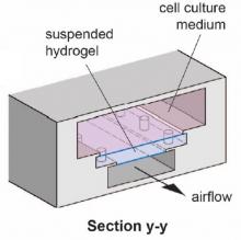
Researchers at the University of Toronto have developed a microfluidic lung airway-on-a-chip device that allows for easy downstream analyses using conventional techniques (e.g. immunohistochemistry, PCR). The device is called E-FLOAT, an acronym standing for “Extractable Floating Liquid gel-based Organ-on-a-Chip for Airway Tissue Modelling”. It consists of three vertically stacked microfluidic compartments; one to contain cell culture media, a second to contain a “floating” hydrogel, and the last through which physiological air can be flowed (Figure 1). Key to the design are certain architectural elements, “microanchors” and a protruding lip, that serve to hold the hydrogel in place in the presence of airflow-induced pressure. Airway epithelial and bronchial smooth muscle cells can be cocultured in the matrix-based hydrogel, which mimics the lamina propria layer existing between the two cell types in bronchioles. Following experimentation, the hydrogel can be easily removed for analysis by various techniques.

Figure 1. E-FLOAT device cross-sections. The device is composed of compartments (left figure) to contain cell culture media, a suspended hydrogel, and airflow. The cell-containing hydrogel provides a barrier between the media and airflow compartments (right figure) and is anchored by “microanchors” and protruding lips. The impact of airborne particles can be assessed using this in vitro lung model.
COMPETITIVE ADVANTAGE
- Off-chip analyses possible with easily extractable gel
- Immunohistochemistry, histology sectioning, scanning electron microscopy, genetic analysis (PCR), ELISA, cell lysis, cell isolation, single-cell RNA sequencing, flow cytometry, matrix stiffness measurements
- Gel mimics the native extracellular matrix
- Gel is able to withstand airflow (~95% RH, and 37 °C)
- Unique geometric features used to prevent gel detachment or leakage during experiments
- Gel remained intact for >96 h with flow rates of 0.6 cm3 s−1 or higher
- Mimics bronchioles, as opposed to stretch-based devices which mimic alveoli
- High manufacturability and arrayability
- 24 usable channels fabricated and ready for cell culture within 6 hours.
APPLICATIONS
- in vitro respiratory research
- Study of airway diseases such as asthma, COPD, cystic fibrosis, and lung cancer,
- Study of disease-causing pollutants
INTELLECTUAL PROPERTY STATUS
- Provisional patent application filed (Sept, 2021)
PROJECT STATUS
Proof-of-concept studies have been conducted. The lung airway-on-a-chip device has been constructed and the cells in the gel layer grown and characterized. The cell type composition closely matched that of normal human lung airways (goblet cells ~10–15%; ciliated epithelial cells ~ 30%) and the cilia length was comparable to those in the native respiratory tract. To demonstrate off-chip analysis, the extractable hydrogel was isolated and characterized using immunohistochemistry, histology sectioning, and scanning electron microscopy.