BACKGROUND
Devices or sensors that can measure cardiomyocyte contractility can provide information on cardiotoxicity or efficacy of drug candidate molecules, in additional to having applications in cardiac research. The most prominent methods of measuring contractility include impedance plates, multielectrode arrays (MEAs), optical imaging methods, and custom techniques that require specialized substrates or methods. These methods are either inaccurate, have low-throughput or require complex set-ups. Alternatively, high frequency ultrasound has been used to determine contractility parameters at the single cell level, but its widespread application has been hindered by poor temporal resolution.
TECHNOLOGY
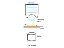
Researchers at the University of Toronto have developed an ultrasound system capable of detecting cardiomyocyte contractility parameters (i.e. beat rate, beat rhythm, force of contraction) at scales ranging from single cells to 3D microtissue constructs. Placement of the ultrasound probe above the sample (Figure 1) allows for simultaneous optical and acoustic imaging. Shifts in acoustic wavelengths induced by the beating cardiomyocytes can be converted to temporally dependent height changes which yield the beat rate and rhythm. The force of contraction can also be measured after determination of the elastic modulus using traction force microscopy. Key to the whole system is its rapid acquisition speed (1000 fps) that allows for resolution of single mouse cardiomyocytes of up to 9 Hz.
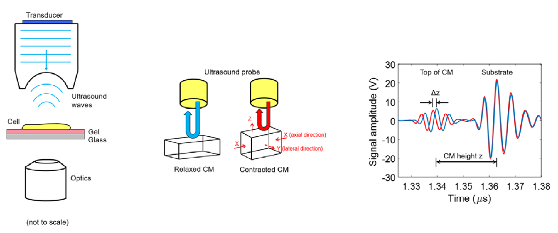
Figure 1. Ultrasound system for measuring cell contractility. The transducer is placed on the top of optical imaging system (left). Contraction of the cardiomyocytes (CMs) lead to changes in height (center) which translate into shift in the received ultrasound wavelengths (right).
COMPETITIVE ADVANTAGE
- Non-invasively and label-free monitoring of cardiomyocyte beat and contractile function
- Measures beat rate, rhythm, and contraction strain
- Unprecedented temporal resolution
- High resolution of 1000 fps resolved the beat profile of single mouse CMs paced at up to 9 Hz
- Amenable to standard well plates and custom fabricated devices
- Single cell to 3D-tissue measurements possible
- Validated
- Contraction force measurements similar to five other techniques
APPLICATIONS
- Drug screening
- Toxicology evaluation
- Cardiac cell therapeutic monitoring
- Modelling disease physiology
INTELLECTUAL PROPERTY STATUS
- National Phase: CA, US (April 2020)
PROJECT STATUS
Proof-of-concept studies have been conducted. The ultrasound system was built on top of a commercial light microscope to enable simultaneous acoustic and optical imaging. Contractile properties of single cells (cardiomyocytes from CD1 male mice) were measured using ultrasound and benchmarked to optical techniques. The technique was also validated on 3D cardiac spheroids, where contractile changes were measured in the presence of two model drugs (i.e. epinephrine and dofetilide).